The Visual Pathways:
Roadmaps and impacts following brain injury
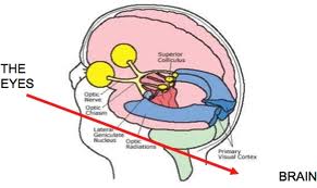 The visual process is so tremendously complex. SO much neurology is involved. And therefore, so much is exposed, following brain injury.
It is not possible to anticipate all of the problems which may result, following a brain insult (such as a stroke or a trauma). However, the array of possible problems may come to light by studying a quick overview of the Visual Pathways.
This summary was presented, following a question which arose from a Medical Resident studying to be an Internist, who received his first exposure to Behavioral Optometry watching my interview. He asks:
I watched part of your interview and I was really impressed at your concept of holistic eye care. This sounds very novel in the field of eye care.
Do you also work with post-stroke patients who have issues with sight due to involvement of their optic track?
My response includes an outline of the visual pathways…
Oculomotor impacts of brain injury
- These signals vary in quality (for example, providing a steady movement, for “smooth pursuit,” or providing a rapid movement or eye-jump, for “saccades”).
- They also vary in the muscle pairings: For example, to look from left to right with both eyes as a team, we require paired constriction of the left medial rectus with the right lateral rectus. But to look from far to near, or to converge the eyes, requires pairing of the left medial rectus and the right medial rectus.
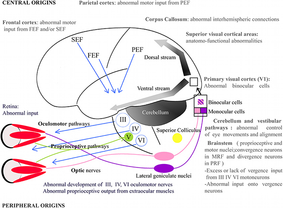
“Eyesight” & steady fixation
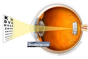 Eye “sight” is merely a matter of “resolution” at the central part of the vision. Eye-movement skills do play some role in that: If the eyes cannot maintain steady fixation, it is more difficult to sustain an image on the center of the eye (fovea) while viewing detail.
Visual information processing post-brain injury
Visual discrimination/ recognition, the ability to “make sense” of things, is largely impacted post-ABI, as this entails visual information PROCESSING. This is activity taking place in the cerebral cortex. It does not only take place in the occipital lobe of the cortex, which is a common misconception among lay-people. True, the visual data which reaches the cortex goes to the striate cortex (IVth layer) of the occipital lobe first. But next the data branches upwards and downwards for gross and fine processing of the “receptive fields.” (Receptive fields are areas over which data is gathered from the retina.) 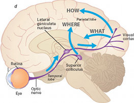 Next, associative cortices support the processing of the data, as we make sense of the world:
- Data are carried towards the parietal lobe for spatial processing, “WHERE is it?”
- Data are carried towards the temporal lobe for identification, “WHAT is it?”
- The frontal lobe also plays a role in planning, such as visual-motor planning- more like, “HOW do I get there, and what should I manage first, etc…”
Visual Pathways
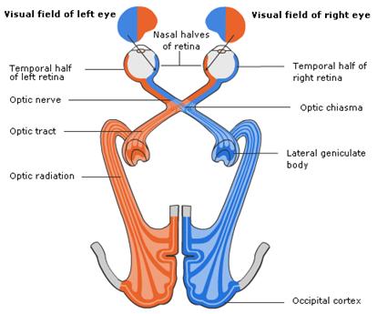
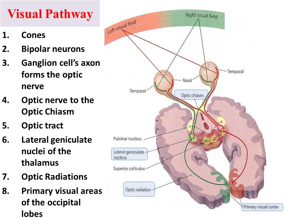
Once light strikes the retina, 90% of the data it is carried via the OPTIC NERVE and RE-SORTED along the OPTIC TRACT on its way to the THALAMUS (Lateral Geniculate Nucleus of the thalamus). Within the thalamus, the neurons synapse before the upper neurons carry the data to the VISUAL CORTEX.
Top-down neurological connections >> bottom-up!
HOWEVER, for every ONE neural pathway carrying data UP to the cortex, there are NINE pathways FROM the CORTEX, TO the THALAMUS, which FILTER the data. Basically, we have 9 times as many “traffic-cops” which limit and filter the data available to the visual system. THEREFORE, when there is damage to the cerebral cortex, some of those Top-down FILTERS are disrupted, and the individual has a great deal of trouble making sense of the overwhelming amount of data available via the visual process.
This is also something that we treat in a Vision Rehabilitation program.
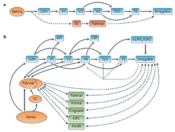
a) 90% of data from the retina goes through the LGN of the thalamus, and onto the visual cortex, then onto associated cortices. 10% bypasses the cortex and goes to Superior colliculus in the brainstem.
b) Diagram shows inter-cortical associations as well as two-way communications between the higher centers of the cortex and the integration centers in the thalamus (pulvinar nucleus), as well as those between the cortex and amygdala (emotional centers), also feeding back to the thalamus. There are many more top-down than bottom-up pathways in the Visual Process.
Sub-cortical visual pathway and Blindsight
Other links of interest
I have written a little blog piece on the eye/brain connection, which you might wish to read. The follow-up piece has some fascinating links on the use of vision in non-sighted individuals as well.


Great information Dr. Slotnick – especially on how the neurology and symptoms are related.
Thank you, Dr. Simonson!
It is the dorsal pathway that is the WHERE, and the ventral is WHAT. You have them reversed.
Thank you, Deborah. You’re absolutely right. Thanks for the content correction!
Thank you, Dr. Simonson.It is the ” HOW” pathway unfamiliar to me.